Inferior Removal of Dislocated Polymethyl Methacrylate Intraocular Lens and Scleral Refixation in Glaucomatous Eyes
Video 1: The inserted IOL was fixed in the sclera using Yamane’s method. The details of the proposed methodology are presented.
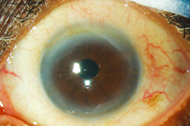
Effect of Different Preoperative Intraocular Pressures on the Prognosis of Traumatic Cyclodialysis Cleft Associated with Lens Subluxation
Peer-reviewed videos describing the effect of different preoperative pressures on the prognosis of traumatic cyclodialysis cleft associated with lens subluxation.
Video 1: The capsular bag is implanted to support the zonules, and a foldable intraocular lens is inserted into the capsule. After zonular dialysis, a 13-mm Morcher Type 2 L CTR, MCTR, with two perforations is pre-placed into the ciliary sulcus with 10-0 polypropylene, and then the end of the Prolene suture is placed on the sclera and it is tied 1 mm behind the edge of the surgical
site.
Video 2: The location of a small hole is pushed to the maximum height of the fissure, which causes the detached ciliary body to be reconnected to the scleral spur via direct mechanical packaging. Furthermore, the cyclodialysis cleft closure was confirmed by gonioscopy at the end of surgery.
An iPhone 7 clipped with a macro lens aligned along the smartphone camera was used in a standard slit-lamp setup, where the light source was projected onto the anterior segment structure of interest, and then the lens-clipped smartphone was slowly advanced towards the subject’s eye. At a distance of 3 to 5 cm from the ocular surface, a fine focus of the area of interest was obtained using the auto focus option. Multiple pictures, magnified or otherwise, and/or video recordings were obtained.
ESM2
Disc and retinal findings were observed using iPhone XS Max, and it was discovered to offer much better quality images with a wider field of view.
ESM3
Visualization of peripapillary area and retinal nerve fiber layers is aided in a clearer and replicable manner. Video clips were advantageous in terms of quality over the standstill images.
Outcomes of adjusted trabeculotomy in cases with juvenile glaucoma
Video showing excerpts of the surgical technique implemented in the study
Combined Visco-Trab operation: A dual filtration pathway for management of advanced glaucoma—midterm results
The Video presenting Visco-Trab operation combined with clear cornea temporal phacoemulsification and in-the-bag foldable intraocular lens implantation in a patient with advanced primary open-angle glaucoma and posterior subcapsular cataract.Steps of combined Visco-Trab operation include fornix-based conjunctival flap and subconjunctival Mitomycin C 0.3 mg for 3 min followed by through wash with BSS. A 4x4mm lamellar scleral flap (1/3-1/2 of thickness) extending 1mm in clear cornea. Deep scleral flap dissection 0.5 mm inside edge, leaving thin scleral over choroid. Exposure & deroofing of Schlemm’s canal. When the glaucoma procedure is combined with phacoemulsification, once the canal is deroofed, the surgeon moves to the temporal side to do the cataract operation through a clear-cornea incision. At the end of the phaco procedure, the AC is reformed and the corneal wound is secured by one 10/0 Nylon suture and the surgeon reverts back to upper position to complete the filtering operation. Excision of deep scleral flap creating a scleral lake. Dilation of SC on either side with sodium hyaluronate. Penetrating trabeculectomy (1×2 mm) and wide peripheral iridectomy were done followed by water tight closure of lamellar scleral flap and conjunctival flap.
Other Procedures for Pediatric Glaucoma Surgery: New Devices and Techniques
Video 1 : Bioniko eye model for simulation of angle surgery. The TRAB™360 surgical technique and GATT are performed in this model
Video 2 : TRAB™360 surgical technique . The instrument is used to pierce the trabecular meshwork; the inner filament is threaded 180° in Schlemm’s canal and then unroofed. The same procedure is performed for the remaining 180°.
Video 3 : Gonioscopic-assisted transluminal trabeculotomy (GATT) with the iTrack™ catheter . The catheter is grasped and directed to the opening in Schlemm’s canal. The catheter is then fed into the canal and then Schlemm’s canal is unroofed.
Nd:YAG Laser for Ahmed Tube Shunt Blockage in Patients Implanted with Boston Type I Keratoprosthesis
Video : Case 2 video: piston-like movement of the micro air bubble in the tube was observed.
Surgical Considerations in
Children with Corneal Opacities and Cataracts
Video 1 : Surgical video with narration demonstrating optical iridectomy in Peters anomaly
Video 2 : Ab externo 360° trabeculotomy , lysis of iridocorneal adhesions, and temporal optical sector iridectomy using illuminated catheter in a child with Peters anomaly. (Courtesy of Alana L. Grajewski, MD)
Case 47: Zone I/II Open Globe Repair with Post-Operative Elevated Intraocular Pressure
Video 1 : Case 47_Zone I-II Blunt Open Globe Injury
Video 1 : Blunt dissection of the correct sub-Tenon’s plane may be started with scissors and completed with two squint hooks inserted back-to-back in this pocket and pulled gently apart. (Courtesy of Cecilia Fenerty, MD, FRCOphth and Tanya Karaconji, MD, FRANZCO)
Video 2 : Method of inserting a tube into the anterior chamber using a blunt-tipped cannula. The tip of the cannula is firmly engaged onto the cut bevel of the tube and gently inserted through the scleral tunnel taking the tube with it. (Courtesy of Cecilia Fenerty, MD, FRCOphth and Tanya Karaconji, MD, FRANZCO)
Video 3 : A long tunnel in the patient’s native sclera created using a small mini-crescent blade (1.25 mm in width) that can be used to tunnel in the sclera up to about 2 mm posterior to the limbus, then completing the entry into the eye with a 25-gauge needle. (Courtesy of James D. Brandt, MD)
Video 4 : Insertion of the tube of a Baerveldt glaucoma drainage device in an eye with aniridia . The lens is slightly displaced to the nasal side, and insertion of the tube at a tangent avoids contact with the lens. The anterior chamber infusion maintains the anterior chamber and constant intraocular pressure throughout the procedure. (Courtesy of Cecilia Fenerty, MD, FRCOphth and Tanya Karaconji, MD, FRANZCO)
Bleb wall recession technique to repair giant bleb formation after Ahmed Glaucoma Valve implantation: a case report (1 Videos)
Video S1. Surgical video of the bleb wall recession technique.
Repair of angle recession prevents pupillary capture of intrasclerally fixed intraocular lenses
Video : This video demonstrates the representative technique for suturing the iris root
Cyclodestruction
Video 1 : Transscleral cyclophotocoagulation . The footplate of the laser probe is placed at the limbus, while laser is activated by a foot pedal and applied to the ciliary body. A pair of fine-toothed forceps helps to rotate the eye during the procedure
Video 2 : Endoscopic cyclophotocoagulation . The tip of the endoprobe is positioned from the ciliary body such that about four to five ciliary processes are visible on the screen at any given time. Laser is applied to the ciliary processes until shrinkage and whitening occur without rupture of ciliary processes or bubble formation.
Nasal goniotomy technique, shown using locking forceps at the superior and inferior limbus, and a Barkan goniotomy lens modified with the addition of a handle, sitting on a cushion of viscoelastic applied to the cornea. A 25-gauge needle is used to enter the anterior chamber and to make a cleft to either side, while the assistant stabilizes the globe and rotates it to expose additional angle. Note that the needle entry is closed with a single buried10-0 polyglactin suture. (Courtesy of Sharon F. Freedman, MD)
Video 2
Nasal goniotomy technique without traction suture or assistant using modified Swan-Jacob lens. The anterior chamber is maintained with viscoelastic and a 25-gauge needle. (Courtesy of Alana Grajewski, MD)
Video 3
Nasal goniotomy without an assistant using a standard Swan-Jacob lens and a 25-gauge needle on viscoelastic. (Courtesy of and narrated by Elizabeth Hodapp, MD)
Electronic supplementary material
Intermediate results of phaco-endocycloplasty in an exclusive cohort of angle closure glaucoma: potential for change
ESM 1
Video that demonstrates technique of phaco-endocycloplasty and ECPL in pseudoexfoliation
ESM 2
Figure demonstrating quiet eye at 1-week post-phaco-ECPL with unscathed conjunctiva